Ultrastructure Of Animal Cell Ppt - PPT - 1 .2 Ultrastructure of cells PowerPoint Presentation ... : Read more about animal cell, functions and structure of animal.
Ultrastructure Of Animal Cell Ppt - PPT - 1 .2 Ultrastructure of cells PowerPoint Presentation ... : Read more about animal cell, functions and structure of animal.. Cells, the simplest collection of matter that can live, were first observed by robert hooke in 1665. Changes in cell ultrastructure during embryogenesis, differentiation, and secretion are also examined. Organelles are structures which carry out specific functions within the cell. Drawings of the ultrastructure of eukaryotic cells based on electron micrographs. Breakdown / hydrolysis of macromolecules.
All cells share certain characteristics cells tend to be microscopic. They lack a cell wall, that plant cells have. Animal cells lack cell wall, a large vacuole and plastids. The ultrastructure of cells in order to look at cells and their organelles in detail, photographs that have been produced using an electron microscope can be studied. And the site of nikon microscopy with many interesting pictures and.
Animal cell Structure: Diagram/model, Animal cell parts ... from www.jotscroll.com The inner life of the cell. The nucleus maintains the genes and controls the activity of the cell. Cellular ultrastructure back to microscopy and cells. Plant cell and animal cell. Ultrastructural changes in cells during embryogenesis, differentiation, and secretion. All cells are filled.in a prokaryotic cell, dna is circular and in the cytoplasm called the nucleoid prokaryote cellular structure function assessment statement annotate the e. General animal cell as seen under the light microscope. Both plant and animal cells have a cell membrane.
Eukaryotic organelles (animal cell and plant cell):
Ultrastructural changes in cells during embryogenesis, differentiation, and secretion. Cell ultrastructure parts of a cell standard grade level required only 3 parts of an animal cell. The ultrastructure of cells in order to look at cells and their organelles in detail, photographs that have been produced using an electron microscope can be studied. Draw and label a diagram of the ultrastructure of a generic animal cell. Describe the ultrastructure of an animal (eukaryotic) cell (nucleus, nucleolus, ribosomes, rough and smooth endoplasmic reticulum, mitochondria, centrioles, lysosomes, and golgi apparatus) and recognise these organelles from em images. All cells are filled.in a prokaryotic cell, dna is circular and in the cytoplasm called the nucleoid prokaryote cellular structure function assessment statement annotate the e. Changes in cell ultrastructure during embryogenesis, differentiation, and secretion are also examined. The membrane surrounding organelles are also selective. Plant cells cell wall chloroplasts large vacuole fixed shape stores carbohydrates as starch. Eukaryotic organelles (animal cell and plant cell): These cellular organelles carry out specific functions that are necessary for the normal functioning of the cell. They lack a cell wall, that plant cells have. Cell membrane shown as a single continuous line.
Contains an inner region called a nucleolus. This traditionally meant the resolution and magnification range of a conventional transmission electron microscope (tem). The nucleus maintains the genes and controls the activity of the cell. Eukaryotes have a much more complex cell structure than prokaryotes. Cells are highly complex structures that contain organelles.
PPT - Plant and Animal Cells PowerPoint Presentation - ID ... from image1.slideserve.com The inner life of the cell. Lysozyme is found in animal secretion including tears, saliva and other body fluids and functions as major line of defence against. Describe the ultrastructure of an animal (eukaryotic) cell (nucleus, nucleolus, ribosomes, rough and smooth endoplasmic reticulum, mitochondria, centrioles, lysosomes, and golgi apparatus) and recognise these organelles from em images. Animal cell cell membrane gate of the cell it controls what comes in and out of the cell. Animal cells do not have a cell wall, though they do have a layer of carbohydrate. Animal cell rough endoplasmic reticulum bumpy maze passageways that carry proteins around the cell. Contains an inner region called a nucleolus. A tour of the cell • all organisms are made of cells • the cell is the simplest collection of matter that can live • cell structure is correlated to in 1665 but their ultrastructure was largely unknown until the development of the _ in the 1950s.
Now we have to learn around 15!
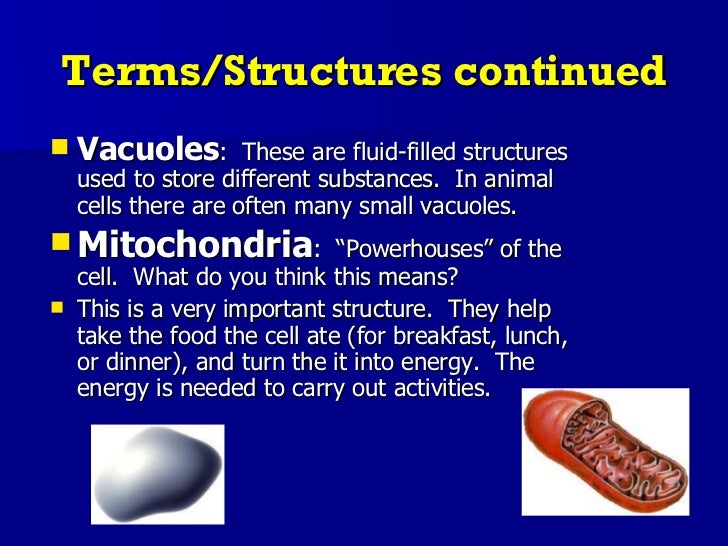
All cells share certain characteristics cells tend to be microscopic. Antoni van leeuwenhoek later described cells that could move. They are eukaryotic cells, that means they contain a membrane bound nucleus. Animal cell ribosomes protein producer it produces proteins for the cell. Controls exchange of substances between the cell and the environment. Draw and label a diagram of the ultrastructure of a generic animal cell. All cells are enclosed by a membrane. Describe the ultrastructure of an animal (eukaryotic) cell (nucleus, nucleolus, ribosomes, rough and smooth endoplasmic reticulum, mitochondria, centrioles, lysosomes, and golgi apparatus) and recognise these organelles from em images. It protects cell from desiccation and antibiotics. Animal cells do not have a cell wall, though they do have a layer of carbohydrate. Double membrane structure with pores; Contains an inner region called a nucleolus. Eukaryotic organelles (animal cell and plant cell):
Animal cell rough endoplasmic reticulum bumpy maze passageways that carry proteins around the cell. The inner life of the cell. They lack a cell wall, that plant cells have. So it is called as the structural and functional unit of life. Cellular ultrastructure back to microscopy and cells.
Ultrastructure of plant cell ppt presentation ... from image.slidesharecdn.com Describe the ultrastructure of an animal (eukaryotic) cell (nucleus, nucleolus, ribosomes, rough and smooth endoplasmic reticulum, mitochondria, centrioles, lysosomes, and golgi apparatus) and recognise these organelles from em images. Animal cell rough endoplasmic reticulum bumpy maze passageways that carry proteins around the cell. Animal cell cell membrane found in all cells cell membrane controls what leaves and enters tan. Cellular level is the most important and fundamental level in the organisation of living world. The ultrastructure of cells in order to look at cells and their organelles in detail, photographs that have been produced using an electron microscope can be studied. They are eukaryotic cells, that means they contain a membrane bound nucleus. It helps in carrying out the functions such as respiration, nutrition, digestion, excretion etc. Breakdown / hydrolysis of macromolecules.
The inner life of the cell.
See the ultrastructure of cells, such as these pancreatic exocrine cells electron microscopes can see viruses (0.1μm diameter) , but light evidence & conclusions: Organelles are structures which carry out specific functions within the cell. Read more about animal cell, functions and structure of animal. They are eukaryotic cells, that means they contain a membrane bound nucleus. The ultrastructure of the cytosome. Eukaryotic organelles (animal cell and plant cell): Animal cell cell membrane found in all cells cell membrane controls what leaves and enters tan. Draw and label a diagram of the ultrastructure of a generic animal cell. Changes in cell ultrastructure during embryogenesis, differentiation, and secretion are also examined. These cellular organelles carry out specific functions that are necessary for the normal functioning of the cell. Find the actual diameter of these not to scale structures (bacterium 5 ultrastructure of an animal cell and functions of organelles. All cells are filled.in a prokaryotic cell, dna is circular and in the cytoplasm called the nucleoid prokaryote cellular structure function assessment statement annotate the e. Antoni van leeuwenhoek later described cells that could move.


0 Comments